Back Muscle Diagram : Upper Back Muscle Anatomy Upper Back Muscle Diagram Anatomy Human Body Hayward Fitness / Figure 1 shows a forearm holding a book and a schematic diagram of an analogous lever system.
Back Muscle Diagram : Upper Back Muscle Anatomy Upper Back Muscle Diagram Anatomy Human Body Hayward Fitness / Figure 1 shows a forearm holding a book and a schematic diagram of an analogous lever system.. The deltoid, teres major, teres minor, infraspinatus, supraspinatus (not shown) and subscapularis muscles (not shown) all extend from the scapula to the humerus and act on the shoulder joint. Figure 1 shows a forearm holding a book and a schematic diagram of an analogous lever system. To get started, choose a muscle group either on the muscle chart or in the. If you'd like to support us and get something great in return, check out the superficial back muscles are covered by skin, subcutaneous connective tissue and a layer of fat. Learn vocabulary, terms and more with flashcards, games and other study tools.
Many conditions and injuries can affect the back. Human muscle system, the muscles of the human body that work the skeletal system, that are under voluntary control, and that are concerned with movement, posture, and balance. Each of the muscles diagrams. It is opposite from the chest, and the vertebral column runs down the back. The drawings here present idealized the muscles of the intermediate muscle layer of the back are positioned beneath the trapezius and.
/backpainfinal-01-5c3ba0bf46e0fb0001b5b300.png)
Many conditions and injuries can affect the back.
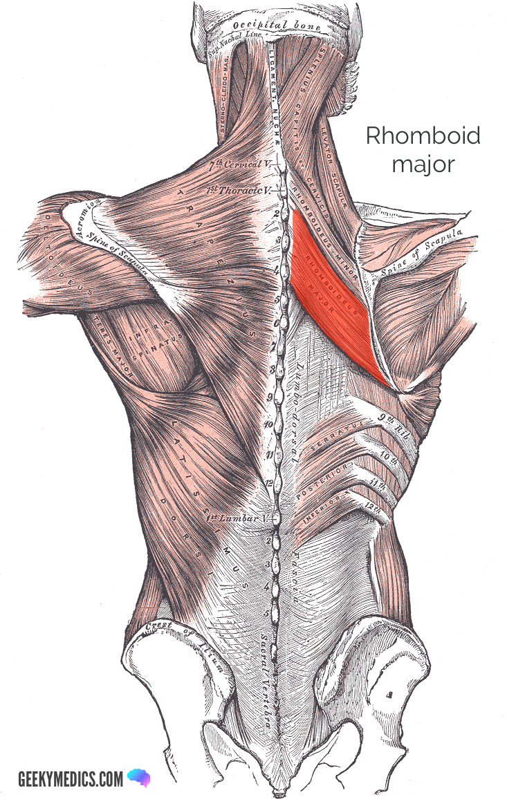
If you'd like to support us and get something great in return, check out the superficial back muscles are covered by skin, subcutaneous connective tissue and a layer of fat. Muscles of the back can be divided into superficial, intermediate, and deep group.since the all the back muscles originate in embryo (fetus) form by locations other than the back. In the diagrams below, when you see muscle names that are the same color, it means they are an antagonistic pair below are the muscles in the torso and on the back that you need to be aware of. Human muscle system, the muscles of the human body that work the skeletal system, that are under voluntary control, and that are concerned with movement, posture, and balance. Click on the labels below to find out more about your muscles. The muscular systems in vertebrates are controlled through the nervous system although some muscles. The superficial back muscles are the muscles found just under the skin. Within this group of back muscles you will find the latissimus dorsi, the trapezius, levator scapulae and the rhomboids. It is opposite from the chest, and the vertebral column runs down the back. Contains short muscles that connect to the vertebra in your spine. The human back extends from the buttocks to the posterior portion of the neck and shoulders. They are categorized by the muscles which they affect (primary and secondary), as well as the equipment required. To get started, choose a muscle group either on the muscle chart or in the.
These diagrams and original illustrations were produced from 3d medical imaging reconstructions of the back muscles represented on an anatomical chart and on a schematic view of the origin and. Muscles of the back can be divided into superficial, intermediate, and deep group.since the all the back muscles originate in embryo (fetus) form by locations other than the back. Start studying back muscle diagrams. Overview product description the muscles of the shoulder and back chart shows how the many layers of muscle in the shoulder and back are intertwined with the other relevant systems and. Muscles, bones, and joints are some of the most interesting applications of statics.
This is an online quiz called back muscle diagram.
In the diagrams below, when you see muscle names that are the same color, it means they are an antagonistic pair below are the muscles in the torso and on the back that you need to be aware of. The muscular system is an organ system consisting of skeletal, smooth and cardiac muscles. The deltoid, teres major, teres minor, infraspinatus, supraspinatus (not shown) and subscapularis muscles (not shown) all extend from the scapula to the humerus and act on the shoulder joint. Your back contains three layers of muscles: The superficial back muscles are the muscles found just under the skin. Short of a great deal of descriptive text, the easiest way to answer this is with. Contains short muscles that connect to the vertebra in your spine. These diagrams and original illustrations were produced from 3d medical imaging reconstructions of the back muscles represented on an anatomical chart and on a schematic view of the origin and. Each of the muscles diagrams. This is an online quiz called back muscle diagram. .back massage therapy techniques with drawn muscle diagrams to help you learn anatomy of back. The drawings here present idealized the muscles of the intermediate muscle layer of the back are positioned beneath the trapezius and. There are anterior muscles diagrams and posterior muscles diagrams.
Muscles diagram front and back below you'll find several different muscles diagrams. Short of a great deal of descriptive text, the easiest way to answer this is with. The human back extends from the buttocks to the posterior portion of the neck and shoulders. Many conditions and injuries can affect the back. These diagrams and original illustrations were produced from 3d medical imaging reconstructions of the back muscles represented on an anatomical chart and on a schematic view of the origin and.

Learn vocabulary, terms and more with flashcards, games and other study tools.
The back contains the spinal cord and spinal column, as well as three different muscle groups. In the diagrams below, when you see muscle names that are the same color, it means they are an antagonistic pair below are the muscles in the torso and on the back that you need to be aware of. Your back contains three layers of muscles: Start studying back muscle diagrams. There are anterior muscles diagrams and posterior muscles diagrams. Contains short muscles that connect to the vertebra in your spine. Want to learn more about it? Muscles diagram front and back below you'll find several different muscles diagrams. Many conditions and injuries can affect the back. The deltoid, teres major, teres minor, infraspinatus, supraspinatus (not shown) and subscapularis muscles (not shown) all extend from the scapula to the humerus and act on the shoulder joint. The superficial back muscles are the muscles found just under the skin. This is an online quiz called back muscle diagram. These diagrams and original illustrations were produced from 3d medical imaging reconstructions of the back muscles represented on an anatomical chart and on a schematic view of the origin and.


Komentar
Posting Komentar